


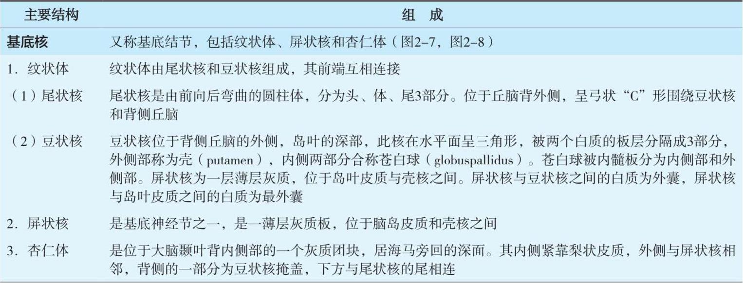
大脑半球皮质称大脑灰质,表面下的白质称髓质。髓质深部的灰质核团为基底神经核。端脑的内腔为侧脑室(表2-4,图2-7,图2-8)。
表2-4 端脑内部主要结构
续表

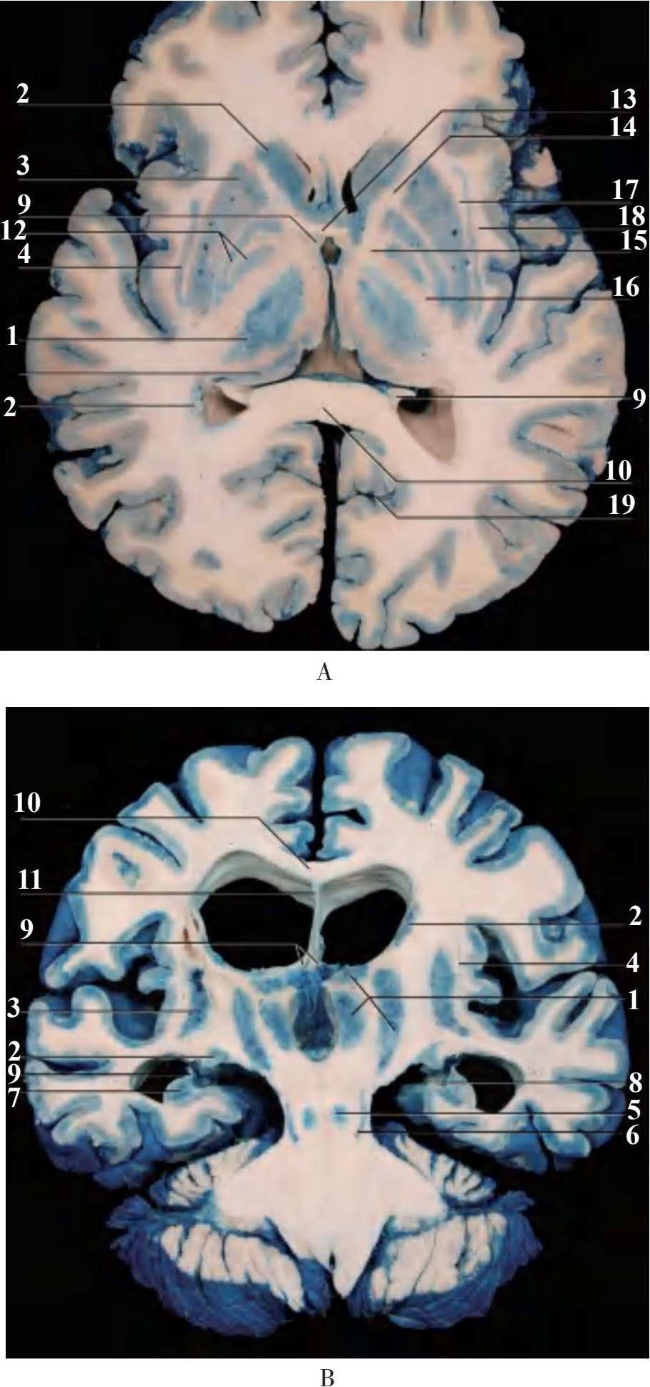
1.丘脑 thalamus;2.豆状核 lentiform nucleus;3.纹状核壳 putamen;4.屏状核 claust rum;5.红核 red nucleus;6.黑质 substantia nigra;7.海马 hippocampus;8.齿状回 dentate gyrus;9.穹隆 fornix;10.胼胝体 corpus callosum;11.透明隔transparent septum;12.苍白球 globus pallidus;13.前连合 anterior commissure;14.内囊前肢 anterior limb of internal capsule;15.内囊膝 genu of internal capsule;16.内囊后肢 posterior limb of internal capsule;17.外囊 external capsule;18.最外囊exterme capsule;19.距状沟 calcarine sulcus
图2-7 端脑内部主要结构
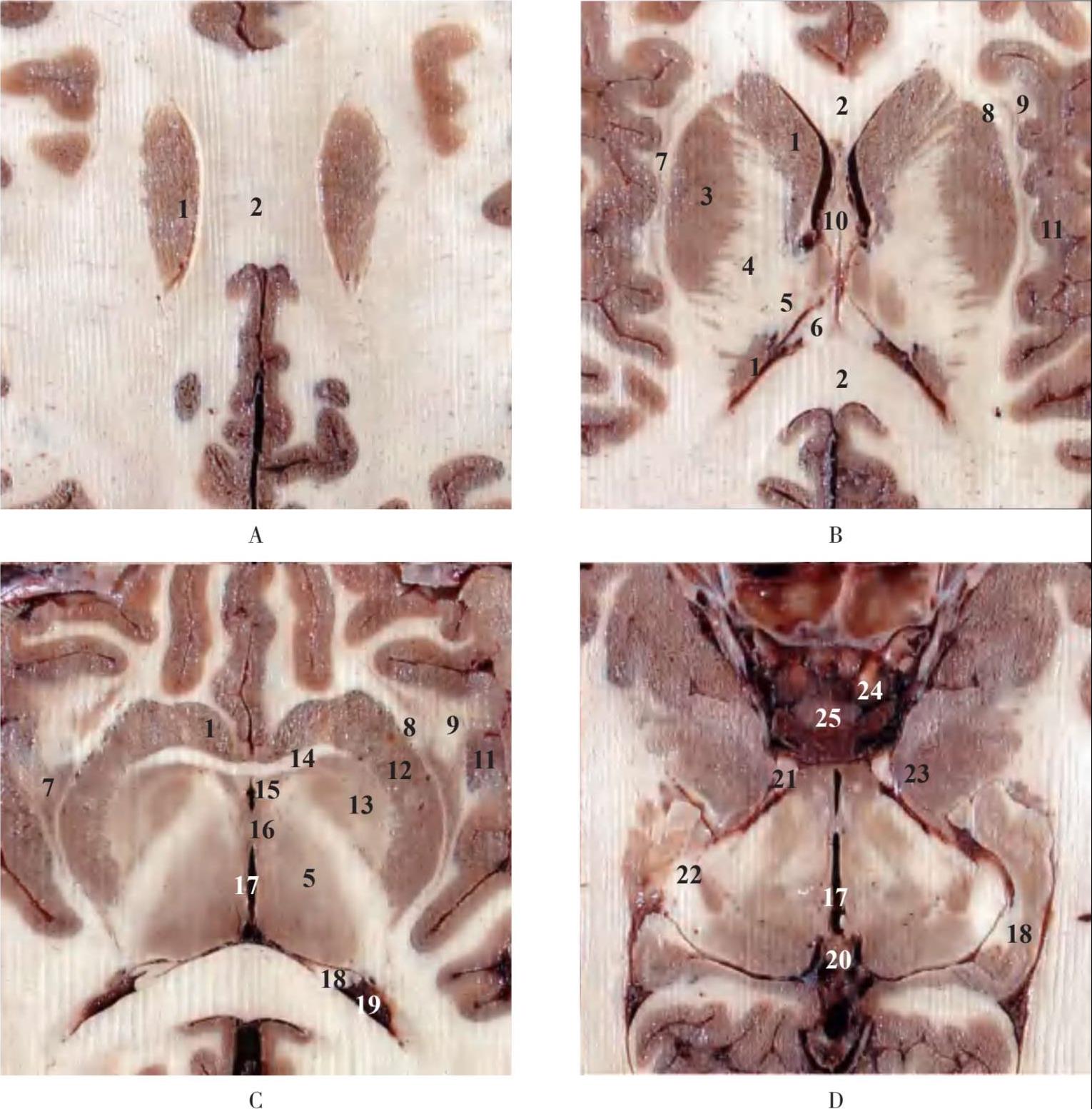
1.尾状核 caudate nucleus;2.胼胝体 corpus callosum;3.豆状核 lentiform nucleus;4.内囊 internal capsule;5.丘脑thalamus;6.穹隆 fornix;7.屏状核 claus trum;8.外囊 external capsule;9.最外囊 exterme capsule;10.透明隔 transparent septum;11.岛叶 insular lobe;12.纹状核壳 putamen;13.苍白球 globus pallidus;14.前连合 anterior commissure;15.穹隆柱 column of fornix;16.中间块 massa intermedia;17.第三脑室 third ventricle;18.海马 hippocampus;19.第四脑室 fourth ventricle;20.松果体 pineal body;21.动眼神经 oculomotor nerve;22.外侧膝状体 lateral geniculate body;23.钩回 uncus;24.颈内动脉 internal carotid artery;25.垂体 pituitary gland
图2-8 水平断层示脑内主要结构
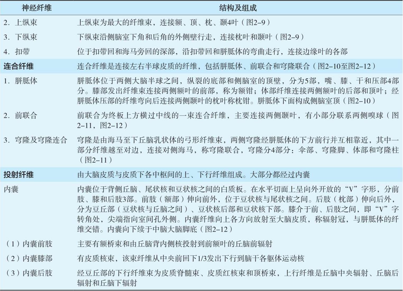
大脑半球的髓质主要由联系皮质各部和皮质下结构的神经纤维组成,可分成3类:联络纤维(association fibers)(图2-9)、连合纤维(commissural fibers)(图2-10,图2-11)、投射纤维(projection fibers)(表2-5,图2-12)。
表2-5 大脑的纤维

续表
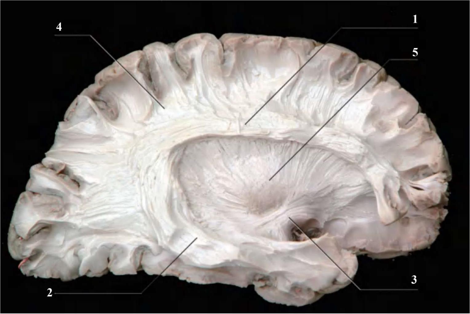
1.上纵束 fasciculus longitudinalis superior;2.下纵束 fasciculus longitudinalis inferior;3.钩束 fasciculus uncinatus;4.大脑弓状纤维 fibrae arcuatae cerebri;5.豆状核 lentiform nucleus
图2-9 联络纤维
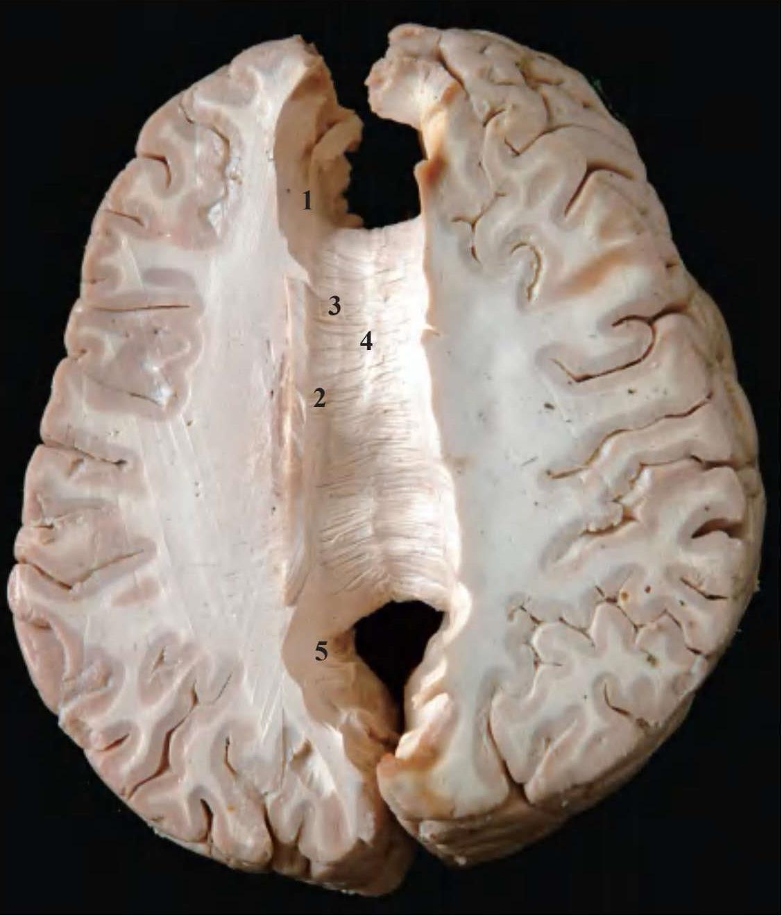
1.额钳 frontal forceps;2.胼胝体辐射 radiatio corporis callosita;3.内侧纵纹 medial longitudinal stria;4.外侧纵纹 lateral longitudinal stria;5.枕钳 occipital forceps
图2-10 连合纤维示胼胝体
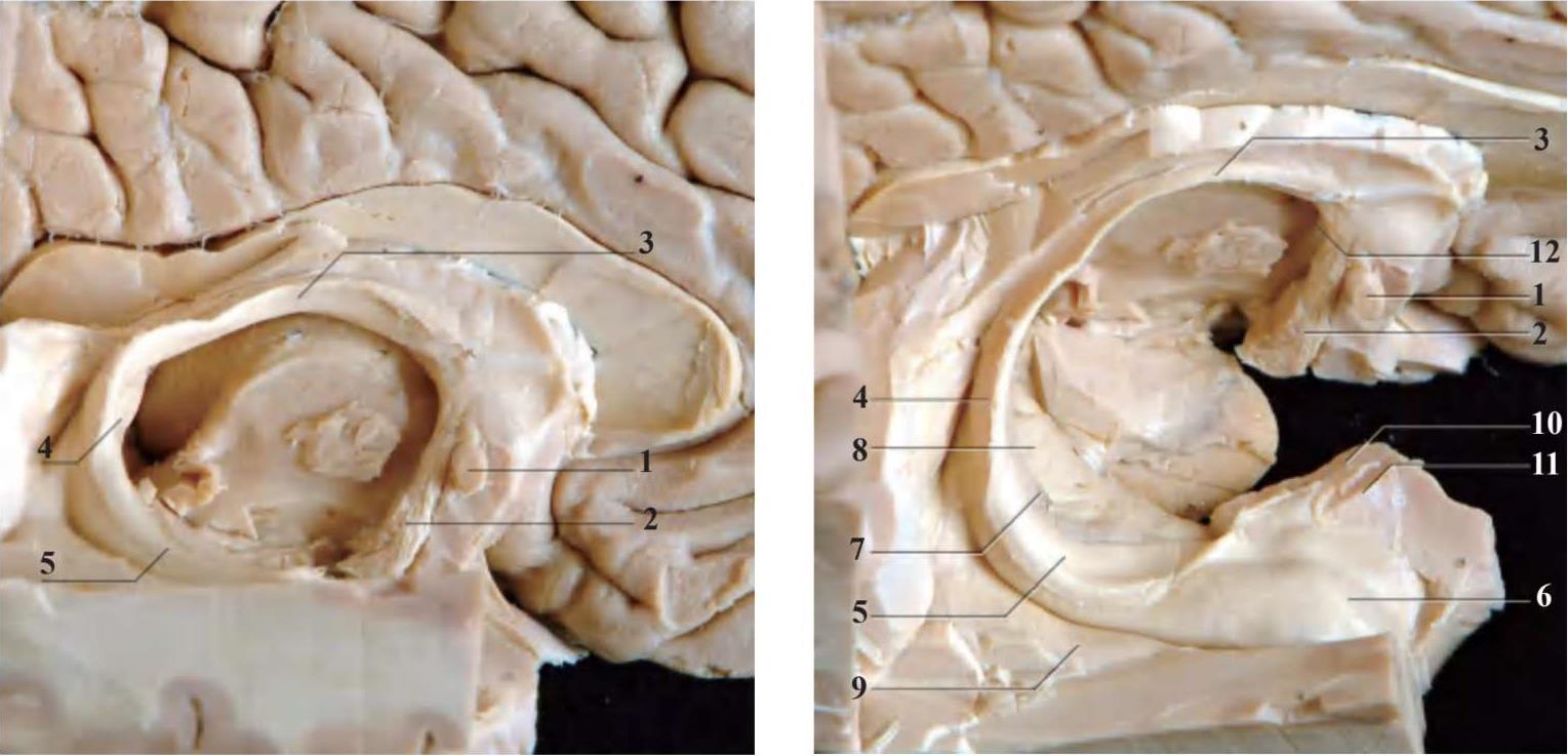
1.前连合 anterior commissure;2.穹隆柱 column of fornix;3.穹隆体 body of fornix;4.穹隆脚 crus of fornix;5.穹隆伞fimbria-fornix;6.海马头 head of hippocampus;7.齿状回 dentate gyrus;8.海马旁回 parahippocampal gyrus;9.侧副三角collateral trigone;10.钩回 uncus;11.杏仁体 amygdaloid body;12.第三脑室 third ventricle
图2-11 联合纤维
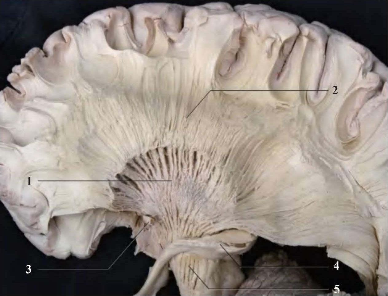
1.内囊 internal capsule;2.辐射冠 corona radiata;3.前连合 anterior commissure;4.视束 optic tract;5.锥体束 pyramidal tract
图2-12 投射纤维
表2-6 大脑主要皮质功能定位
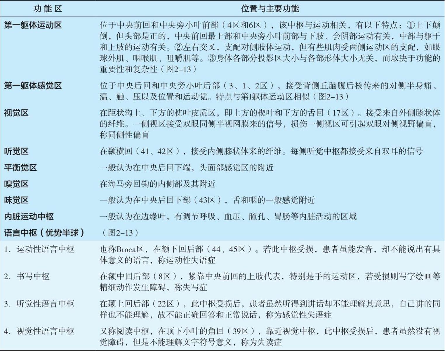
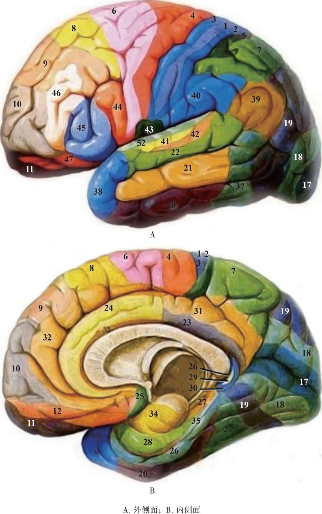
1~3.体感皮层主要体感觉区 primary somatosensory cortex;4.主要运动区 primary motor cortex;5.体感联络皮层 somatosensory association cortex;6.前运动皮层和辅助运动区 premotor cortex and supplementary motor cortex(secondary motor cortex);7.视觉运动协同区 visuo-motor coordination;8.额叶眼动区 frontal eye fields;9.后外侧前额叶皮层 posterior lateral prefrontal cortex;10.额极区(上额回和中额回最前侧)anterior prefrontal cortex(most rostral part of superior and middle frontal gyrus);11.额眶区(眶回、直回及上额回前侧)orbitofrontal area(orbital and rectus gyri,plus part of the rostral part of the superior frontal gyrus);12.额眶区(上额回及下前回之前)orbitofrontal area(used to be part of BA11,refers to the area between the superior frontal gyrus and the inferior rostral sulcus);13.岛皮层 insular cortex;17.初级视觉区 primary visual cortex(V1);18.次级视觉区 secondary visual cortex(V2);19.视觉联络区 associative visual cortex(V3~V5);20.颞下回 inferior temporal gyrus;21.颞中回 middle temporal gyrus;22.颞上回,其前侧部分属于韦尼克区 superior temporal gyrus,a part of Wernicke’s area;23.下后扣带皮层 ventral posterior cingulate cortex;24.下前扣带皮层 ventral anterior cingulate cortex;25.膝下皮层 ectosplenial portion of the retrosplenial region of the cerebral cortex;26.压外区 ectosplenial area;27.梨状皮质piriform cortex;28.后内嗅皮层 ventral entorhinal cortex;29.压后扣带皮层 retrosplenial cingulate cortex;30.扣带皮层的一部分part of cingulate cortex;31.上后扣带皮层 dorsal posterior cingulate cortex;32.上前扣带皮层 dorsal anterior cingulate cortex;33.扣带前回 anterior cingulate gyrus;34.前扣带皮层 dorsal cingulate cortex;35.旁嗅皮层 perirhinal cortex;36.海马旁皮层 parahippocampal cortex;37.梭状回 fusiform gyrus;38.颞极区 temporopolar area;39.角回 angular gyrus,韦尼克区(Wernicke’s area);40.缘上回 supramarginal gyrus,韦尼克区(Wernicke’s area)的一部分;41、42.初级听皮层和听觉联合皮层 primary auditory cortex and auditory association cortices;43.中央下区 subcentral area;44.三角部 pars triangularis,布洛卡区(Broca’s area)的一部分,语言运动区;45.岛盖部 pars opercularis,布洛卡区(Broca’s area)的一部分,语言运动区;46.上外额叶皮层 dorsolateral prefrontal cortex;47.下额叶皮层 pars orbitalis;48.下脚后区 retrosubicular area;52.岛旁区 parasubicular area
图2-13 大脑Brodmann分区
(引用自The Neocortex of macaca mulatta)
(冯展鹏 王军 张沛东;标本制作:石瑾 骆承恩)
[1]栾丽菊,刘丰春.中央前、后回在矢状断面上的定位[J].中国临床解剖学杂志,2004,22(2):179-182.
[2]李七渝,张绍祥,刘正津,等.大脑横断面解剖与MRI对照研究[J].中国医学影像技术,2005,21(4):639-642.
[3]刘丰春,栾丽菊.端脑髓型对脑回定位的影像解剖学研究[J].中国临床解剖学杂志,2003,21(3):291-292.
[4]李雪鹏,李少华,张剑凯,等.端脑横切面上髓突与中央前后回的对应关系[J].中国临床解剖学杂志,2006,24(6):645-647.
[5]张华,周庭永,钱学华,等.人丘脑及相关重要结构的断面解剖学研究[J].中国临床解剖学杂志,2012,30(4):393-397.
[6]李雪鹏,张剑凯,李少华,等.额叶脑回的横断层定位[J].解剖与临床,2006,11(6):375-377.
[7]周健,栾国明.颞叶癫痫的外科相关解剖[J].立体定向和功能性神经外科杂志,2001,14(1):54-57.
[8]朱镛连.大脑两半球的解剖、功能与障碍[J].中国康复理论与实践,2011,17(6):598-600.
[9]陈一勇,尹维刚,林荣,等.成人岛叶纤维连接的显示和分析[J].解剖学杂志,2014,37(5):661-663,675.
[10]冯三平,冯继,周益民,等.大脑中动脉与岛叶的显微解剖[J].中华神经外科杂志,2009,25(1):76-78.
[11]GAREY L J.Brodmann’s Localisation in the Cerebral Cortex[M].New York:Springer,2006.
[12]YUSTE R,Church G M.The new century of the brain[J].Scientific American,2014,310(3):38-45.
[13]SPORNS O.Networks of the Brain[M].London:MIT Press,2010.
[14]DAMASIO H.Neural basis of language disorders// R.Chapey,Language intervention strategies in adult aphasia.4th edition[M].Baltimore:Williams & Wilkins,2001.
[15]VARNAVAS O G,GRAND W.The insular codex:morphological and vascular anatomic characteristics[J].Neurosurgery,1999,44(1):127-136.
[16]TANRIOVER N,RHOTON A L Jr,KAWASHIMA M,et a1.Microsurgical anatomy of the insula and the sylvian fissure[J].J Neurosurg,2004,100(5):891-922.
[17]RHOTON A L Jr.The cerebrum anatomy[J].Neurosurgery,2007,61(1):37-118.
[18]BIEGA T J,LONSER R R,BUTMAN J A.Differential cortical thickness across the central sulcus:a method for identifying the central SUICUS in the presence of mass effect and vasogenic edema[J].AJNR,2006,27(7):1450-1453.