
下载掌阅APP,畅读海量书库
立即打开


 Isolation of the Islets of Langerhans for Transplantation
D. R. Thomas et al.
Editor’s Note
This paper describes a method for isolating rat islets of Langerhans as a potential transplant source for diabetes. Attempts to treat diabetes by transplanting a whole pancreas had met with limited success, and efforts to isolate the component insulin-producing islets of Langerhans were hampered by methodological problems and low viability. The revised method, by D. R. Thomas and colleagues at Sheffield in England, includes digestion, washing and separation steps and yielded large numbers of viable islets of Langerhans. Islet cell transplantation is currently being assessed as a potential treatment for type I diabetes mellitus, where the immune system destroys insulin-producing beta cells in the islets of Langerhans. Successful transplants have reduced or removed the need for insulin therapy. 中文
PANCREATIC transplantation with the aim of treating diabetes mellitus has so far met with little success. Of 23 patients thus treated and reported to the Transplant Registry in 1971 1 , 15 died within 3 months and the longest survived one year. One of the major problems has been to overcome pancreatic exocrine digestion, and pancreatic duct ligation (“Banting pancreas”) before transplantation has been performed. It was shown by Dragstedt 2 , however, that dogs treated in this way often became diabetic or showed a diabetic glucose tolerance test after several months, probably due to fibrosis and consequent ischaemia of the Islets. Transplantation of the whole gland with its vascular supply is a major undertaking and the problems of thrombosis, leakage and digestion, coupled with immunological rejection, have prevented success so far. 中文
Attempts have been made to isolate the Islets of Langerhans from the pancreas in order to study glucose metabolism. A micro-dissection technique was described by Hellerstrom 3 but this was tedious and produced only small numbers of Islet clumps. Subsequently Moskalewski 4 described a method of collagenase digestion which was successful in the rabbit and improved the yield, and Lacy and Kostianovski 5 modified the method for the rat. There were still difficulties in harvesting the liberated Islets, however, even with methods of separation such as zonal centrifugation and density gradients, and there were also problems with viability of the cells. 中文
We have developed a technique by which large numbers of Islets of Langerhans are prepared consistently from the rat pancreas for purposes of transplantation. Viability was confirmed by transplantation. 中文
Initially, albino outbred rats and subsequently young adult inbred hooded rats (strain PVG/C) were used. Following killing by cervical dislocation, the ventral surface of the animal was shaved, prepared with chlorhexidine in spirit and the abdomen opened with a midline incision. Using a dissecting microscope, magnification×10, a fine polyethylene catheter (“Intracath”—B. R. Bard, London) was introduced into the common bile duct and secured with a 2-0 linen thread ligature. The lower end of the duct was occluded with an artery forceps just before its entry into the duodenum. The pancreas was distended by injection of 10 ml. Hanks’ solution containing bovine albumen (fraction V), 2 mg·ml. -1 . It was found that after some practice this procedure could be completed within 5 min of the death of the animal. The pancreas was removed, transferred to a glass Petri dish, cut into small pieces with scissors and any excess fat removed. It was then transferred to a tube to which a further 10 ml. of Hanks’ solution was added. The pancreatic tissue sank to the bottom and any remaining fat floated on the surface and was readily aspirated and discarded. The prepared tissue was transferred to a small conical flask together with 2 ml. Hanks’ solution containing glucose 0.6 mg ml. –1 and collagenase type 1 (Sigma Chemicals, London). The stoppered flask was placed in a shaking water bath at 37℃ for about 30 min. The exact time for separation was determined by frequent sampling and examination under the dissecting microscope. 中文
The digested pancreas was transferred to a tube and diluted with further cold Hanks’ solution containing glucose in the same concentration as before and gently centrifuged for 1 min. The supernatant was discarded and fresh medium added, and the resultant suspension filtered into a Petri dish with a blackened base for examination under the dissecting microscope at×10 magnification. 中文
Initially the view was obscured by fine fragments of acinar tissue, which were removed by gently agitating the Petri dish and allowing them to become suspended in the Hanks’ solution, then aspirated and discarded, the Islets remaining on the bottom of the vessel. They could then be seen clearly (Fig. 1) by the aid of dark ground illumination as yellowish white domes and were picked out with a finely drawn Pasteur pipette. Fig. 2 shows the histological appearance of an isolated pancreatic Islet. Its architecture and cells appear normal. 中文
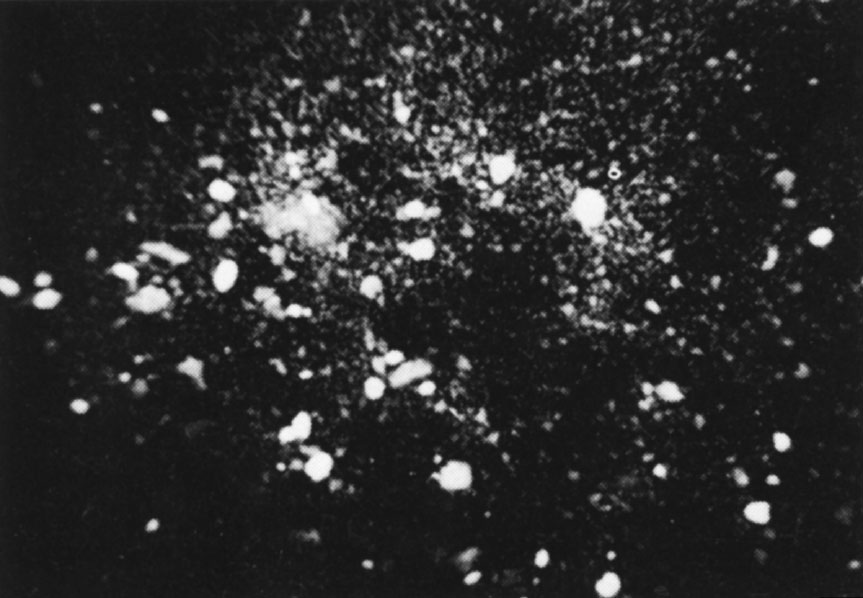
Fig. 1. Islet cell tissue seen after isolation from the pancreas using dark ground illumination. Dissecting microscope, ×5.
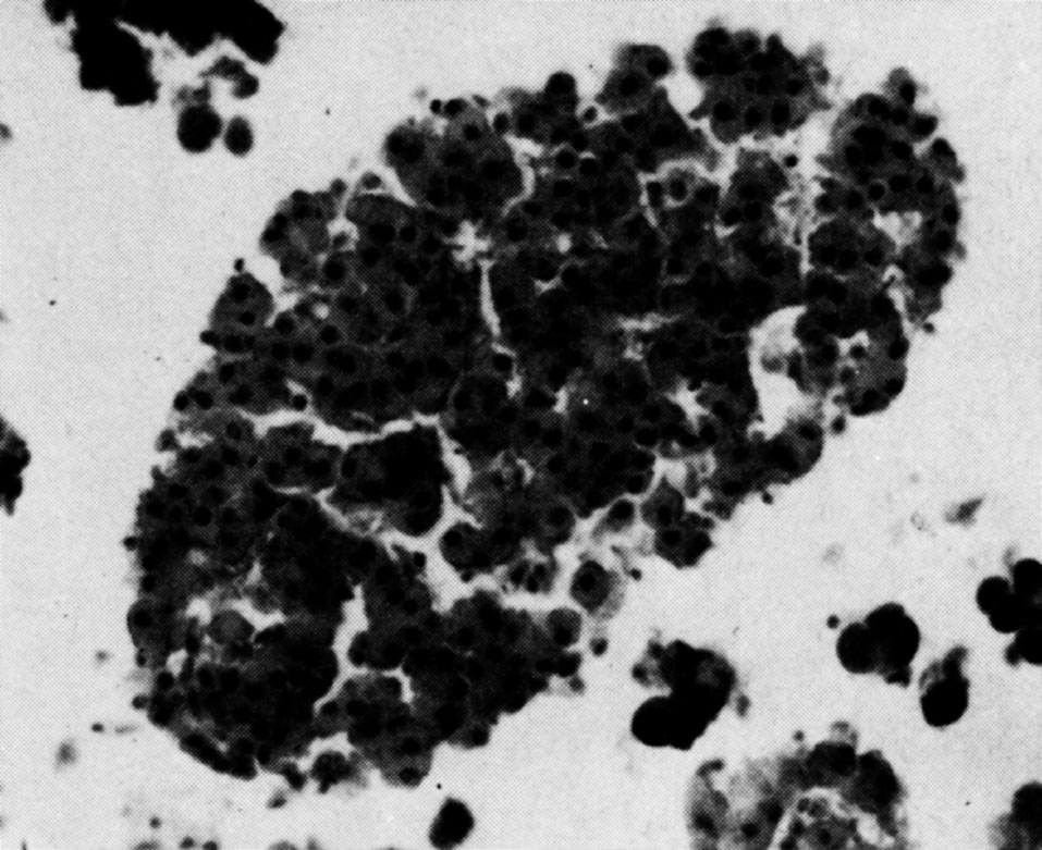
Fig. 2. Isolated pancreatic Islet. Normal architecture and appearance of cells. Haematoxylin and eosin, ×300.
Yields of up to 350 Islets per rat pancreas have been achieved using this method. To obtain larger quantities, rat pancreases have been processed in batches of four. 中文
Viability of the isolated cells was confirmed by transplantation beneath the renal capsule and into the testis of isogeneic rats. The longest period of follow-up was one month, when viable looking Islet cells containing beta cell granules staining with aldehyde-fuchsin were seen. A similar method has been successfully applied in the rabbit. 中文
The relationship of the Islets of Langerhans to diabetes mellitus was established in 1889 by Von Mering and Minkowski 6 , and in 1892 Hedon 7 demonstrated that subcutaneous implantation of a small piece of pancreas could delay the appearance of diabetes in an animal that had undergone pancreatectomy. The concept of Islet cell grafting appears to be neglected in the extensive literature on pancreatic transplantation, although one early report suggests that transplanted Islets may modify alloxan diabetes in the rat 8 . 中文
We have now established a successful method of isolation of Islets of Langerhans in an animal strain in which inbred immunologically isogeneic lines are available, making possible transplantation studies uncomplicated by rejection problems. These cells can be grafted and survive for appreciable periods of time as shown by the viability studies. Histological and functional studies of Islet cell transplantation will be reported elsewhere. 中文
We acknowledge a grant from the Endowment Fund of the United Sheffield Hospitals and from the Medical Research Council. We thank Dr. Laurence Henry for his help with the histology and Mr A. Tunstill and his staff for photography. 中文
( 242 , 258-260; 1973)
D. R. Thomas, M. Fox and A. A. Grieve
Royal Hospital, Sheffield
Received October 12, 1972. Requests for reprints to M. F.
References: